

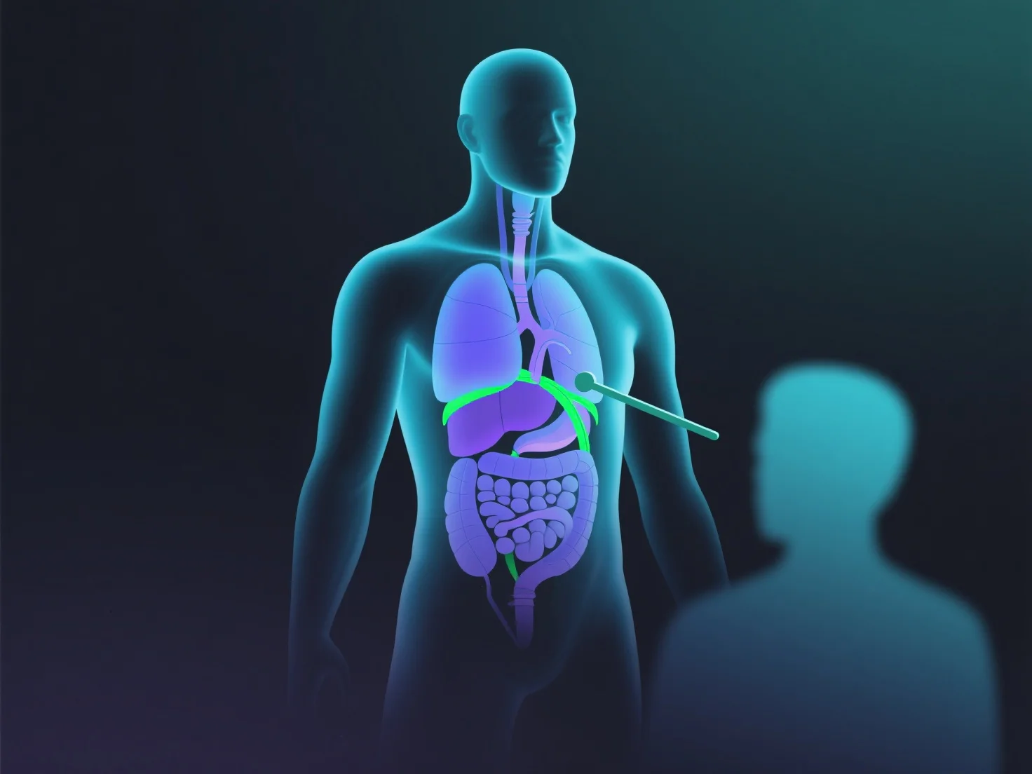
3D modeling enhances the teaching of anatomy in medical education by providing interactive, detailed 3D representations of human structures that students can manipulate and explore.
- **Improves visualization**: It helps learners better understand complex anatomical relationships—such as organ spatial arrangement, tissue layers, or nerve pathways—compared to 2D diagrams or static models. - **Enables virtual dissection**: Students can zoom in on small structures (e.g., blood vessels, nerve endings) or rotate models for multi-angle views, deepening spatial anatomy comprehension. - **Complements traditional methods**: Integrating 3D models into lectures or labs supports cadaver studies, making abstract concepts more concrete and engaging.
This approach strengthens student learning by bridging gaps in traditional anatomy education.